
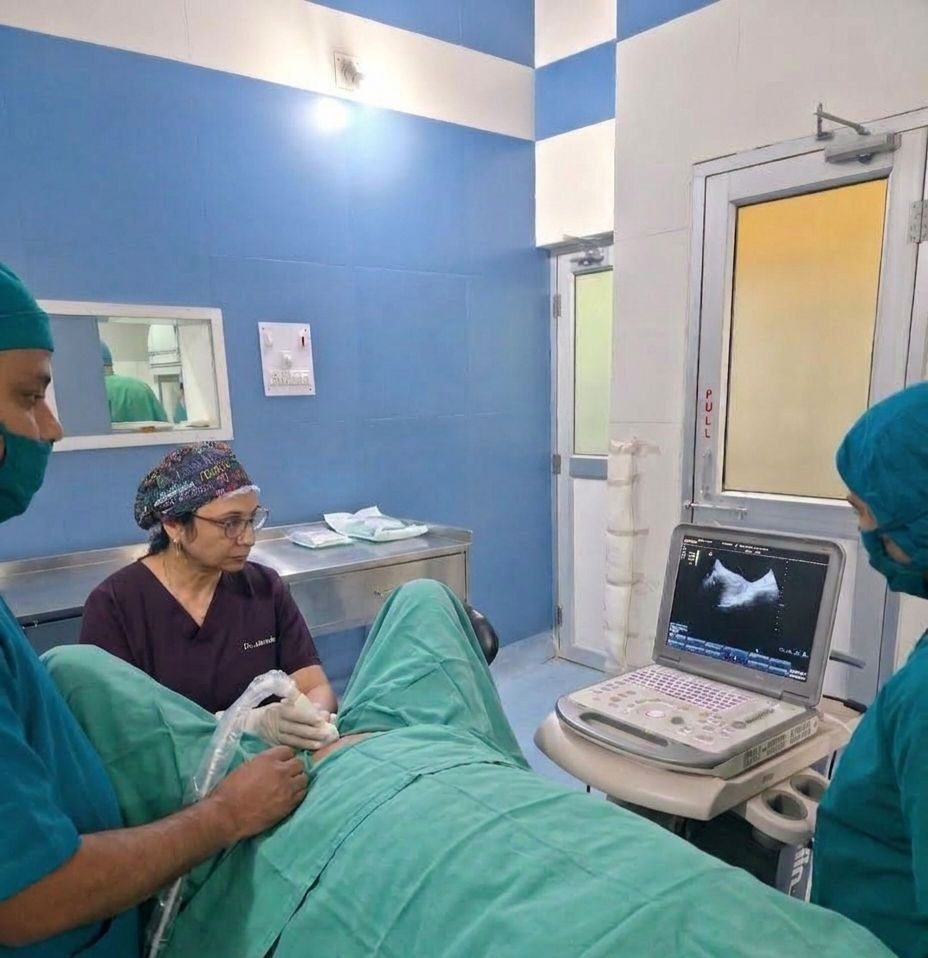
Clinical Pathology
Clinical Pathology — urine, stool and microscopy services in Bokaro Steel City
Clinical pathology is the laboratory’s oldest and most human section: urine under the microscope, stool examination, semen analysis and peripheral smears — the direct, doctor’s-eye tests that machines still cannot fully replace. In an era of expensive panels, these humble tests remain among the most informative in medicine: a urine examination settles the “is this a urine infection?” question in minutes for the price of a snack; a stool test identifies the parasites that quietly anaemise entire families; a semen analysis decides the direction of a couple’s fertility journey. At Surgy Centre’s in-house laboratory, these tests have been processed with care since 1977.
Samples accepted during OPD hours (Monday–Saturday 9 AM–12 PM and 3–6 PM); most reports the same day. Call 88770 88770 / 73710 50505 for guidance.
Urine examination — the two-minute window into your body
What the routine urine test covers
When we rely on it
Collecting a good sample
Midstream, clean, in a sterile container: clean the area, start passing urine, collect mid-flow, cap immediately. Morning samples are best (concentrated); samples should reach the lab within an hour. Bring previous reports for comparison; trends matter.
Stool examination — the unglamorous test that explains so much
Chronic loose motions, recurrent abdominal pain, unexplained anaemia and children who “eat but don’t gain” are daily OPD complaints whose answers often live in a stool report. In our region, worm loads and amoebiasis remain common — treatable conditions that persist untreated because nobody did the simple test. Collection: a clean, dry container, no urine contamination, delivered fresh within an hour; avoid testing during antibiotic courses unless specifically advised.
Semen analysis — the fertility workup’s highest-value test
For couples struggling to conceive, the semen analysis is the single most informative first test — non-invasive, inexpensive, and decisive in direction. It reports count, motility (movement), morphology (shape), volume and liquefaction against WHO reference values. Details of how it fits the wider evaluation are in our Complete Infertility Treatment programme. Practical points: 2–3 days of abstinence before the sample, delivery to the laboratory within an hour (collection rooms are available), no illness or high fever in the preceding weeks (temporarily suppresses counts — retest later), and repeat testing when abnormal, since counts vary naturally between samples. One abnormal report does not define a man; one normal report in a confirmed problem also does not close the question.
Peripheral smear and body-fluid microscopy
Why microscopy still matters in 2026
Automated analysers are wonderful — we run them daily. But machines count; they do not see. The cast morphology that distinguishes kidney diseases, the parasitic ova identified by shape, the smear’s abnormal cell that the analyser flagged only as “abnormal WBC” — these need trained human eyes. Our laboratory keeps both: automation for speed and volume, microscopy for judgement. Every abnormal automated result gets the human review it deserves before reporting.
Give your sample
Laboratory inside Surgy Centre, 225 Cooperative Colony, Bokaro Steel City, Jharkhand 827001. Monday–Saturday 9 AM–12 PM and 3–6 PM; emergency processing for admitted patients 24×7. Phone: 88770 88770 / 73710 50505. Related: All Laboratory Services · Serology · All Treatments.
Urine culture — when the routine test needs a sequel
The urine routine finds the infection’s fingerprint (pus cells, bacteria); the culture names the criminal and its weaknesses. When symptoms say infection — burning, frequency, urgency, fever with flank pain — and especially when they repeat, the culture decides therapy: which bacteria grew, which antibiotics kill it (the sensitivity panel), and therefore which tablet actually works instead of the pharmacy’s guess. The technique is everything: a clean midstream sample (wash, start passing, catch mid-flow) in a sterile container, delivered within the hour — the “morning sample kept overnight on the shelf” culture grows the shelf, not the patient. Who needs cultures: every pregnancy with urinary symptoms (silent infections there threaten the baby), every recurrent infection, every infection that failed the first antibiotic, every catheterised or hospitalised patient with fever. The results rhythm: 24–48 hours for growth, sensitivities the day after — which is why the doctor often starts a sensible antibiotic immediately and phones to switch it when the culture answers. This pairing — routine for speed, culture for precision — is how urinary infections are actually cured rather than merely soothed.
Stool parasites — the burden Bokaro kitchens still carry
The ova-and-parasite examination remains among the most under-requested, highest-yield tests in our region. What it finds: roundworm and hookworm (the anaemia-makers of haematology), giardia and amoeba (the chronic-loose-motion twins), threadworms in scratching children, and the cysts that family cooking chains pass around. Who deserves the test: the child who “eats but doesn’t gain”; the anaemia that iron doesn’t fix; the loose motions that come and go for months; the abdominal pain with the appetite that vanished; the whole family when one member’s report comes back positive (worms are household news, not individual news). The collection rules: fresh sample, clean dry container, no urine mixed, within the hour — and no antibiotics or anti-diarrhoeals masking the picture where avoidable. The prevention that follows treatment: deworming schedules per the family’s exposure, the water and vegetable-washing habits, the hand-to-mouth chain broken at the toddlers. The stool test costs less than a phone recharge and explains more family illness than most families credit — the bias of this page is open and earned.
Semen analysis collection — doing it right the first time
Because the semen analysis decides the direction of an entire fertility workup, its collection standards are strict, and we insist on them kindly. Abstinence: 2–3 days — shorter lowers volume, longer ages the sample. Collection: the private room at our laboratory (preferred) or home collection delivered within 30–60 minutes, body-temperature transported; condoms are forbidden (spermicides kill); coitus interruptus contaminates. Completeness: the whole ejaculate — the first, sperm-rich portion especially; lost fractions distort every number. The illness rule: fever, flu or heavy illness in the prior three months suppresses counts temporarily — test after recovery, not during the aftermath. The repeat rule: one abnormal report earns a second test 2–3 weeks later — counts swing naturally, and decisions are made on patterns. The honesty rule: results go to the couple together; the report that says “low count” says nothing about blame — it says “next step”, and the next step is decided in the consultation, not in the corridor’s imagination.
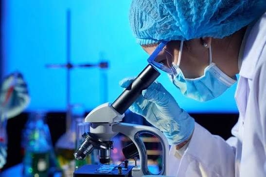
The microscope bench — where machines send their doubts
Every modern laboratory runs on analysers; every good laboratory keeps a microscope bench with senior eyes behind it, and ours is the heart of this section. The analyser flags; the bench decides: the flagged WBC that the smear shows is a leukaemic blast, not a mere count; the “low platelet” that the slide reveals as platelet clumping (a tube artefact — the patient’s platelets were fine all along); the malarial parasite the counter missed; the cast morphology that names the kidney disease; the ova the automated system never learned. The bench’s daily cases: every abnormal CBC smear-reviewed before reporting, every doubtful urine read by eye, every fluid studied cell by cell. Why it matters to you: the difference between a machine’s flag and a cytologist’s verdict is often the difference between a repeat test and a diagnosis — between “come again next week” and “here is what we do now”. Automation bought us speed; the microscope bench kept us honest; both sit in this section, and the patients get the sum.
Body fluids and special microscopy — the chest tube’s story
When fluid collects where it shouldn’t — the chest’s pleural space, the abdomen’s peritoneum — the drained fluid is a document the laboratory reads in full: appearance (clear, cloudy, milky, bloody — each a different story), protein and cell counts (exudate versus transudate — the kidney-liver-infection fork), microscopy and cytology (pus cells and organisms — infection; lymphocytes — tuberculosis’s signature; malignant cells — the finding that changes everything, analysed with the cytology pathway). The same discipline covers joint fluids (the gout crystals, the septic arthritis emergency), wound swabs, and the occasional CSF in urgent collaboration with referral centres. What the family should know: fluid analysis is often the fastest road between “swelling and breathlessness” and “the specific cause with the specific treatment” — and the tests begin the moment the sample leaves the needle, because some of these answers expire in hours.
Need a consultation or second opinion?
Book your appointment online or call us directly — our team will confirm your slot within minutes.